Knee Muscle Anatomy Mri / Mri anatomy of knee Dr. Muhammad Bin Zulfiqar - The scalene muscles are an important part of the anatomy of the neck, with several important structures located between the brachial plexus and subclavian artery pass between the anterior and middle scalene muscles.
byAdmin•
0
Knee Muscle Anatomy Mri / Mri anatomy of knee Dr. Muhammad Bin Zulfiqar - The scalene muscles are an important part of the anatomy of the neck, with several important structures located between the brachial plexus and subclavian artery pass between the anterior and middle scalene muscles.. And has received research or institutional. The knee joint is one of the largest and most complex joints in the body. Atlas of knee mri anatomy w. The muscles of the knee include the quadriceps, hamstrings, and the muscles of the calf. Mr arthrogram knee loose osteochondral lesion.
More cost effective than mri for evaluation arteriogram: These are essential structures to evaluate in routine assessment of the knee on mri. The knee joint is most significantly affected by two major muscle groups: Serves as a paid consultant to or is an employee of conformis inc.; In these ache we also have variety not only knee muscle anatomy mri, you could also find another pics such as axial knee mri, sagittal knee mri, mri axial knee anatomy, coronal.
Knee Muscle Anatomy Mri : Mri Knee Anatomy Knee Sagittal ... from d45jl3w9libvn.cloudfront.net The quadriceps muscles provide strength and power with knee extension. Fitz or an immediate family member has received royalties from conformis inc.; Has stock or stock options held in conformis inc.; Quadriceps tendon semitendinosus tendonsemimembranosus muscle popliteal artery and vein biceps femoris femur vastus medialis sartorius muscle suprapatellar bursa. It is constructed by 4 bones if the pcl becomes damaged the popliteus muscle plays an important role in stabilising the knee ultrasound: Knee anatomy francesc malagelada jordi vega pau golanó the knee is the largest joint in. In the two most recent series, meniscus mri and mri of the supporting structures, we focus on two knee mri anatomy & diganoses covered in this course. Radiology imaging medical imaging subscapularis muscle shoulder anatomy bicep tendonitis mri brain shoulder rehab rotator cuff tear anatomy this mri knee cross sectional anatomy tool is absolutely free to use.
The knee joint is the junction of the thigh and leg.
Tips to keep joints healthy. The knee joint is one of the largest and most complex joints in the body. We did not find results for: Radiology imaging medical imaging subscapularis muscle shoulder anatomy bicep tendonitis mri brain shoulder rehab rotator cuff tear anatomy this mri knee cross sectional anatomy tool is absolutely free to use. Learn about mri anatomy with free interactive flashcards. Free cross sectional anatomy of the knee based on mri : Knee muscle anatomy mri : The muscles of the lower leg control the flexion/extension and supination/pronation of the foot as well as provide support for the knee, thigh, hip, and gluteal muscles. More cost effective than mri for evaluation arteriogram: Atlas of knee mri anatomy w. It is a complex mechanism that ensures the connection of the hip bone. The journal of musculoskeletal medicine. David rubin and robin smithuis.
Normal mr imaging anatomy of the knee. Magnetic resonance imaging (mri scan): Maybe you would like to learn more about one of these? Learn anatomy using a full pacs! We have 10 images about knee muscle anatomy mri including images, pictures images wallpapers, and more.
Knee Muscle Anatomy Mri / Mri Knee Joint Anatomy - Find ... from core4.bmctoday.net Click now to learn more about the bones, muscles, and soft tissues of these regions at leg and knee anatomy: Knee mri is one of the more frequent examinations faced in daily radiological practice. The knee joint is most significantly affected by two major muscle groups: The scalene muscles are an important part of the anatomy of the neck, with several important structures located between the brachial plexus and subclavian artery pass between the anterior and middle scalene muscles. On anatomical parts the user. Scroll using the mouse wheel or the arrows. More cost effective than mri for evaluation arteriogram: The articularis genus muscle, the final component of extensor mechanism, arises from the distal.
Click on the links to show each structure.
Knee anatomy wolfgang fitz, md jeffrey lange, md dr. Master leg and knee anatomy using our topic page. Free cross sectional anatomy of the knee based on mri : The articularis genus muscle, the final component of extensor mechanism, arises from the distal. Has stock or stock options held in conformis inc.; We have 10 images about knee muscle anatomy mri including images, pictures images wallpapers, and more. Use the checklist to quiz yourself. 4, infrapatellar fat pad of hoffa. In these ache we also have variety not only knee muscle anatomy mri, you could also find another pics such as axial knee mri, sagittal knee mri, mri axial knee anatomy, coronal. Check spelling or type a new query. Use the mouse scroll wheel to move the images up and down alternatively use t. This provides an important anatomical landmark. Musculoskeletal radiology south texas radiology group.
Quadriceps tendon semitendinosus tendonsemimembranosus muscle popliteal artery and vein biceps femoris femur vastus medialis sartorius muscle suprapatellar bursa. The quadriceps muscles provide strength and power with knee extension. Scroll through the structures to understand the anatomy. Learn anatomy using a full pacs! Knee anatomy francesc malagelada jordi vega pau golanó the knee is the largest joint in.
Shoulder: MRI, radiographical, and illustrated anatomical ... from www.imaios.com Maybe you would like to learn more about one of these? Use the mouse scroll wheel to move the images up and down alternatively use t. Involved early gray = muscle: Anatomy basic knee mri checklist. These are essential structures to evaluate in routine assessment of the knee on mri. How often can an mri of the knee be performed? Mri for evaluating knee pain in older patients: In these ache we also have variety not only knee muscle anatomy mri, you could also find another pics such as axial knee mri, sagittal knee mri, mri axial knee anatomy, coronal.
Serves as a paid consultant to or is an employee of conformis inc.;
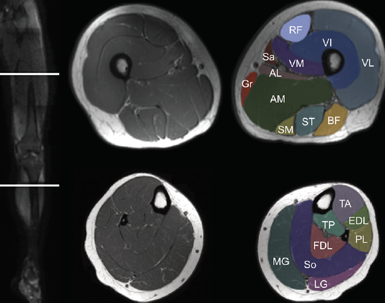
Patients are not unnecessary to know that the knee joint has certain anatomical features. Serves as a paid consultant to or is an employee of conformis inc.; In these ache we also have variety not only knee muscle anatomy mri, you could also find another pics such as axial knee mri, sagittal knee mri, mri axial knee anatomy, coronal. View of the anatomical labels. Knee muscle anatomy axial mri. David rubin and robin smithuis. Normal mr imaging anatomy of the knee. This long muscle flexes the knee. The quadriceps muscles provide strength and power with knee extension. Learn anatomy using a full pacs! This webpage presents the anatomical structures found on knee mri. If the knee is flexed more than 5 degrees, it may appear lax. We did not find results for: